Santiago Ramón y Cajal is an iconic figure in the history of Spanish science. His name is indelibly linked to modern neuroscience thanks to his research on the nervous system, which earned him the Nobel Prize in 1906. However, his legacy is not limited to his scientific discoveries; it also encompasses his role as a teacher who, from the university lecture hall, trained new generations of physicians and researchers.
Throughout his career, the University not only provided him with the material stability necessary to sustain his research and his family, but also a fertile environment for the exchange of ideas and the training of young scientists. The relationship with the University shaped Santiago Ramón y Cajal’s scientific trajectory. It was there that he found the channel for his teaching and research endeavors. It was at the University that he met D. Aureliano Maestre de San Juan, Professor of Normal and Pathological Histology at the Universidad Central de Madrid, who showed him some micrographic preparations that, in their novelty and richness of detail, caused him profound admiration, decisively guiding him toward the study of Histology. It was the University that, as a member of the examination board for the Chair of Anatomy, brought him to Madrid, where he had the fortune of meeting Dr. Simarro. This encounter proved decisive: Simarro showed him nerve cells impregnated with silver, a technique developed by Golgi. This experience led Cajal to redirect his research toward neuroscience, motivating him to use and perfect the silver impregnation technique that Golgi had discovered by chance.
The University allowed him to develop his teaching and formative vocation. Cajal’s bond with the institution began in Zaragoza, where he studied medicine and subsequently served first as an assistant professor and later as an interim professor. His first stable chair came in 1883 at the University of Valencia, as Professor of Descriptive and General Anatomy. From this period, Cajal fondly remembered his “zealous and dedicated students” (“alumnos celosos y aplicados”), among whom he singled out Dr. Manuela Solís, whom he praised for her integrity, honesty, and professional rectitude in the prologue he wrote for her work “Hygiene of Pregnancy and Early Childhood” (“Higiene del embarazo y de la primera infancia”).
After a period as Professor of Histology and Pathological Anatomy at the University of Barcelona, in 1892 he moved to Madrid upon obtaining the chair that had become vacant following the death of D. Aureliano at the Universidad Central. There he would remain until his retirement, establishing himself as a key figure in teaching and research. His work as a professor went beyond the spoken word: with his illustrations, Cajal succeeded in representing with unprecedented precision the complexity of the nervous system and the hypothesis that revolutionized neuroscience — the neuron doctrine.
The impact of Cajal’s illustrations was so remarkable that in 1894, when the polygraphic center was created under the Faculty of Medicine of the Universidad Central, his drawings were selected for the first reproductions. This center, conceived as an auxiliary service, aimed to produce graphic representations intended for use as teaching materials to support the lectures of the various chairs — much like today’s presentations — and to disseminate the discoveries of professors in Histology and Anatomy. The copies produced were to be distributed among the faculties of medicine across the existing university districts in Spain — Madrid, Barcelona, Valencia, Seville, Granada, Santiago de Compostela, Valladolid, and Zaragoza — and, from 1900 onward, also Salamanca, which reopened its medical studies.
The first panels produced by this center are still preserved in the Department of Cell Biology and Histology of the Faculty of Medicine at the Universidad Complutense de Madrid. This department is the direct continuation of the Chair of Histology that Cajal held for 30 years and that served as the cradle of the Spanish Histological School. In addition to these large-format illustrations, only a few additional copies have been located at the universities of Zaragoza and Valencia. These drawings, delineated by Cajal and painted by Ramón Padró y Pedret, a well-known court painter under Alfonso XII, constitute historical and didactic treasures.
The artistic quality of his illustrations was such that they not only facilitated the understanding of microscopic structures but also inspired his students and colleagues. With an extraordinary ability to balance complexity and clarity, Cajal used diagrams, pictograms, and multiple perspectives to formulate hypotheses and explain phenomena more effectively than text alone. His skill as a draftsman allowed him to create visual maps of the brain, the optic system, and various types of cells, revolutionizing the way histology was taught.
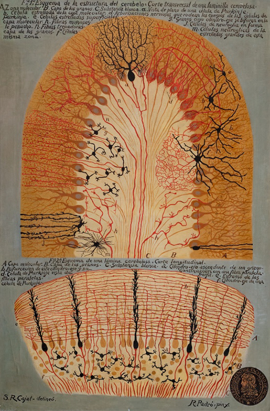
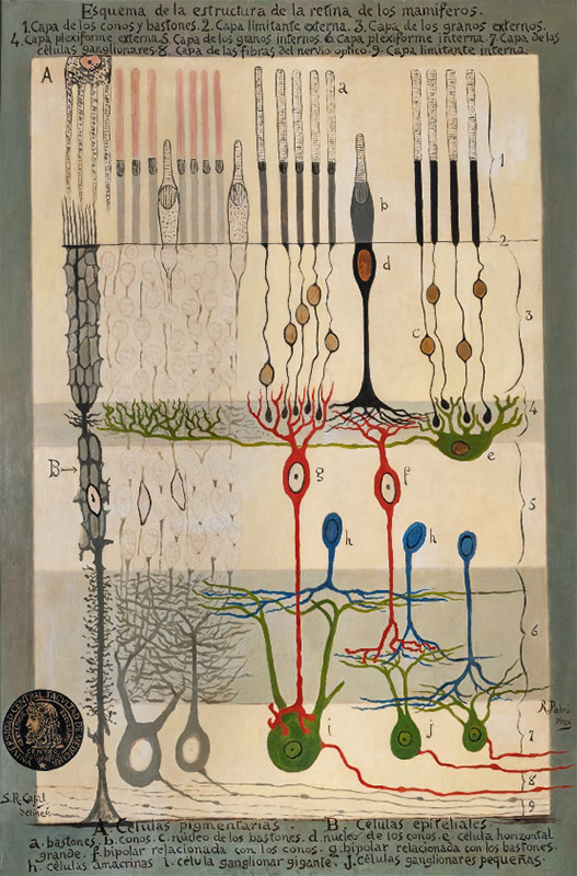
Currently, six of these panels are preserved at the Universidad Complutense, representing various histological structures of the nervous system: the cerebral cortex, the connections of the spinal cord, the structure of the cerebellar cortex (figure 1), the structure of the retina (figure 2), the diagram of the central olfactory apparatus, and the cochlear duct (organ of Corti). Cajal’s studies of the cerebellar cortex and the retina were decisive in demonstrating that neurons are independent cells that contact one another (synapses) — the principles upon which the neuron doctrine is founded. The panel of the cerebellar cortex appears in the background of the portrait painted by the Valencian artist Joaquín Sorolla. In this panel, Cajal schematically shows the arrangement of the different types of neurons and nerve fibers in this structure, compiling in a single drawing the observations obtained through two of the methods most used by Cajal: the Golgi method and his own reduced silver nitrate method.
These illustrations reflect once again Cajal’s effort to provide his students with the resources necessary for a more complete education. Along the same lines, he published two works, “Manual of Normal Histology and Micrographic Technique” (“Manual de Histología Normal y Técnica Micrográfica”) and “Manual of General Pathological Anatomy” (“Manual de Anatomía Patológica General”), which in their time constituted the essential core of knowledge in these disciplines. The Faculty of Medicine, by creating the polygraphic center and choosing his drawings as the first to be reproduced, recognized the value of his initiative and reinforced its commitment to the improvement of medical education.
Cajal’s influence on university teaching extends beyond his scientific legacy: his illustrations, more than mere graphic resources, were a true key tool in medical education. By uniting art and science, he revolutionized the understanding of the nervous system, and his visual method remains a benchmark in teaching and research, demonstrating that science education can be, in addition to rigorous, profoundly aesthetic and inspiring.
Elena Giné Professor at the Universidad Complutense de Madrid I Prize D. Santiago Ramón y Cajal, Professor of Histology: Honorable Mention