With special admiration and respect, this article is dedicated to Professor Isidro Sanchez Garcia (CSIC, Cancer Research Centre, Salamanca). His pioneering research, which has revealed the fundamental connection between stem cells and the origin of leukemia, as well as the causal role of infection in its development, represents the modern continuation of the Cajalian spirit. His unconditional support as a champion of the Salamanca for Cajal and Science initiative is a testament to his commitment. Where Cajal glimpsed the “germinal corpuscles” (“corpusculos germinales”) and the “defensive reaction” (“reaccion defensiva”), the work of Dr. Sanchez Garcia illuminates the molecular mechanisms that govern cancer initiation, carrying the torch of Spanish science toward a new frontier: prevention.
Introduction: The Giant of Neuroscience and the Shadow of His Other Works
The figure of Santiago Ramon y Cajal (1852-1934) stands, with indisputable merit, as the fundamental pillar of modern neuroscience. His monumental body of work, culminating in the Nobel Prize in Physiology or Medicine in 1906 alongside Camillo Golgi, revolutionized our understanding of the nervous system by establishing the neuron doctrine: the theory that the brain is composed of individual, discrete cells. This overwhelming and well-deserved reputation, however, has cast a long shadow over other facets of his scientific genius, eclipsing significant contributions in fields that, at first glance, appear unrelated to his principal line of research, such as his pioneering description of the sarcolemma in the cardiomyocyte. Although Cajal never defined himself as a hematologist in the modern sense of the term, a thorough analysis of his career reveals a body of pioneering and prescient work on inflammation, the morphology of blood cells, and the immune response, which constitutes a substantial contribution to the history of hematology and immunopathology.
This article proposes a chronological and thematic reassessment of Cajal’s non-neuronal work. It will begin with his earliest studies in pathology, which laid the foundations of his biological thinking; it will examine his influential Manual of General Pathological Anatomy as a repository of his ideas on blood pathology; it will analyze his specific publications on the components of blood; and, crucially, it will contextualize his achievements against those of contemporaries such as Paul Ehrlich, the architect of modern hematology. Through this analysis, which will use his meticulous drawings as primary evidence of his morphological genius, it will be demonstrated that the greatness of Cajal does not reside solely in having been the cartographer of the brain, but in having been a universal cell biologist. His capacity to decipher form and function extended to all the “protagonists of life and suffering” (“protagonistas de la vida y del dolor”), including blood cells, revealing a coherence and depth in his thinking that transcends the boundaries of any single discipline.
Figure 1: The Battlefield
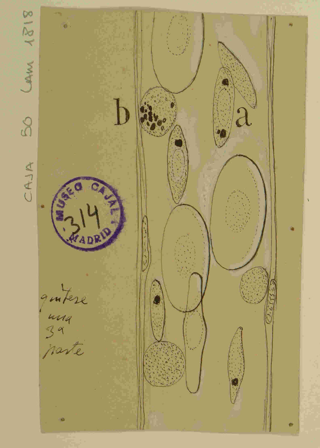
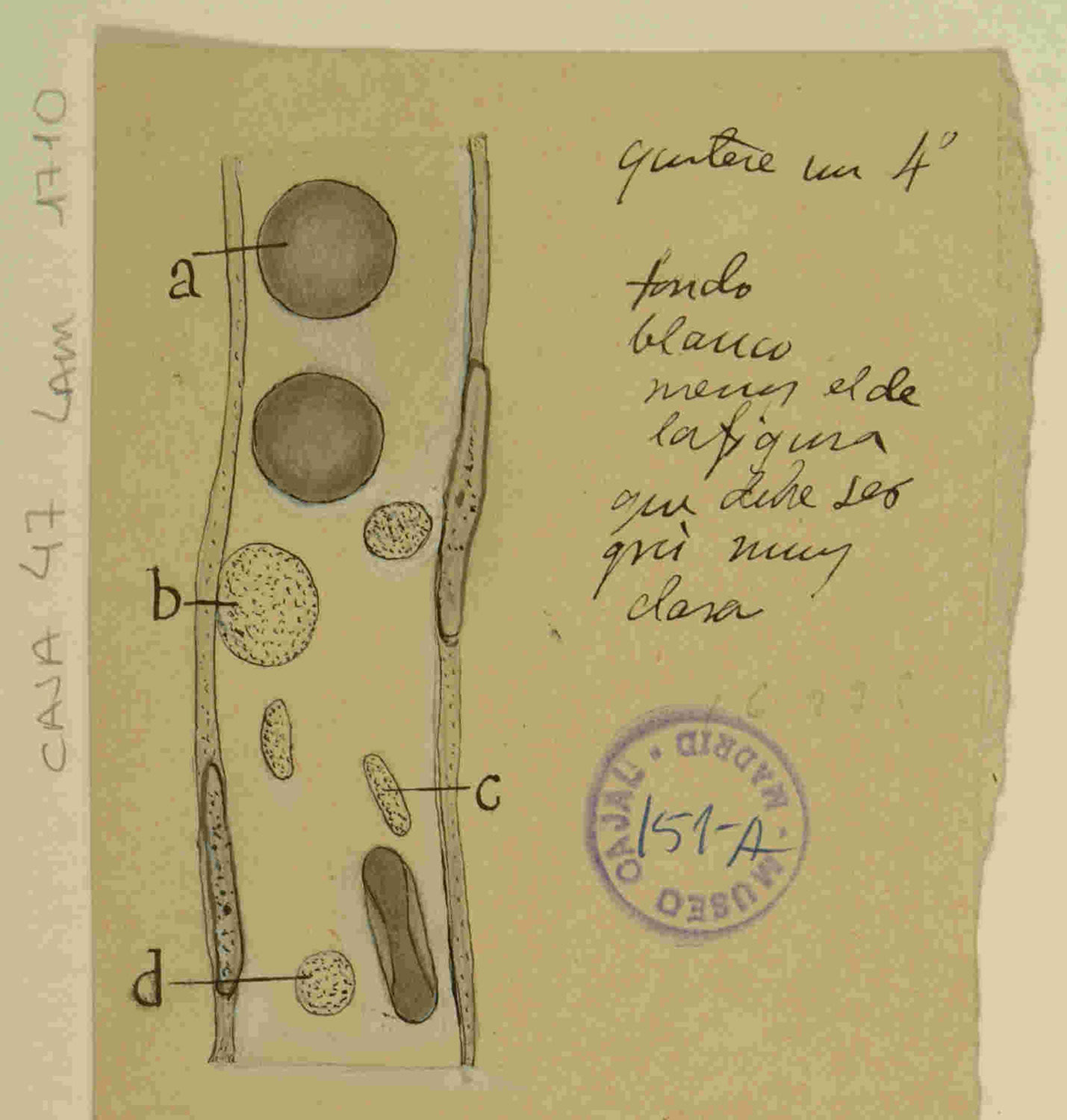
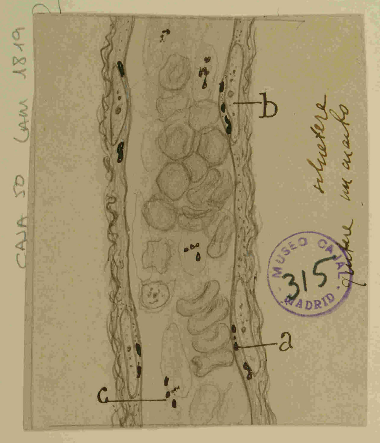
“Frog mesentery capillary” - “Guinea pig mesentery capillary” - “Cat blood vessel” and “Blood capillary”. These drawings depict the settings of Cajal’s earliest discoveries on inflammation. The transparency of the frog mesentery provided him with a unique window to observe in vivo the blood flow and the “pilgrimage” of leukocytes from the vessels into the tissue, the phenomenon that fascinated him and led him to confirm diapedesis.
Chapter 1: The Foundations in Pathology - The Cellular Pilgrimage of Inflammation
1.1: The Doctoral Thesis and the Scientific Debate
The starting point of the illustrious research career of Santiago Ramon y Cajal was not the nervous system, but the microscopic battlefield of inflammation. His doctoral thesis, entitled Pathogenesis of Inflammation (1877), placed him squarely at the epicenter of one of the fiercest debates in nineteenth-century pathology: the origin of pus cells. This work was supervised by Aureliano Maestre de San Juan, a key figure and precursor of modern histology in Spain. The debate was polarized between the influential theory of Rudolf Virchow, who maintained a local origin for inflammatory cells, and the revolutionary hypothesis of Julius Cohnheim (1867), who proposed that leukocytes actively migrated from the blood vessels in a process he termed diapedesis.
Figure 1: The Cellular Pilgrimage
1.2: Experimental Verification and the Philosophy of Drawing
True to his skepticism toward dogma, Cajal addressed the question empirically in his first scientific publication, Experimental Investigations on the Genesis of Inflammation and Especially on Leukocyte Emigration (1880). He replicated Cohnheim’s experimental model, observing inflammation in the frog mesentery, and not only confirmed diapedesis but immortalized it in drawings that are, in themselves, a scientific argument. He himself admitted having spent up to twenty continuous hours “observing the movements of a sluggish leukocyte in its laborious efforts to escape from a blood capillary” (“observando los movimientos de un leucocito perezoso en sus laboriosos esfuerzos por escapar de un capilar sanguineo”). For Cajal, drawing was a “language, an articulation of ideas that allowed thoughts to develop.” His illustrations were not passive transcriptions but active interpretations and syntheses of countless hours of observation, treating his microscopic subjects as animate beings with “motives and yearnings.”
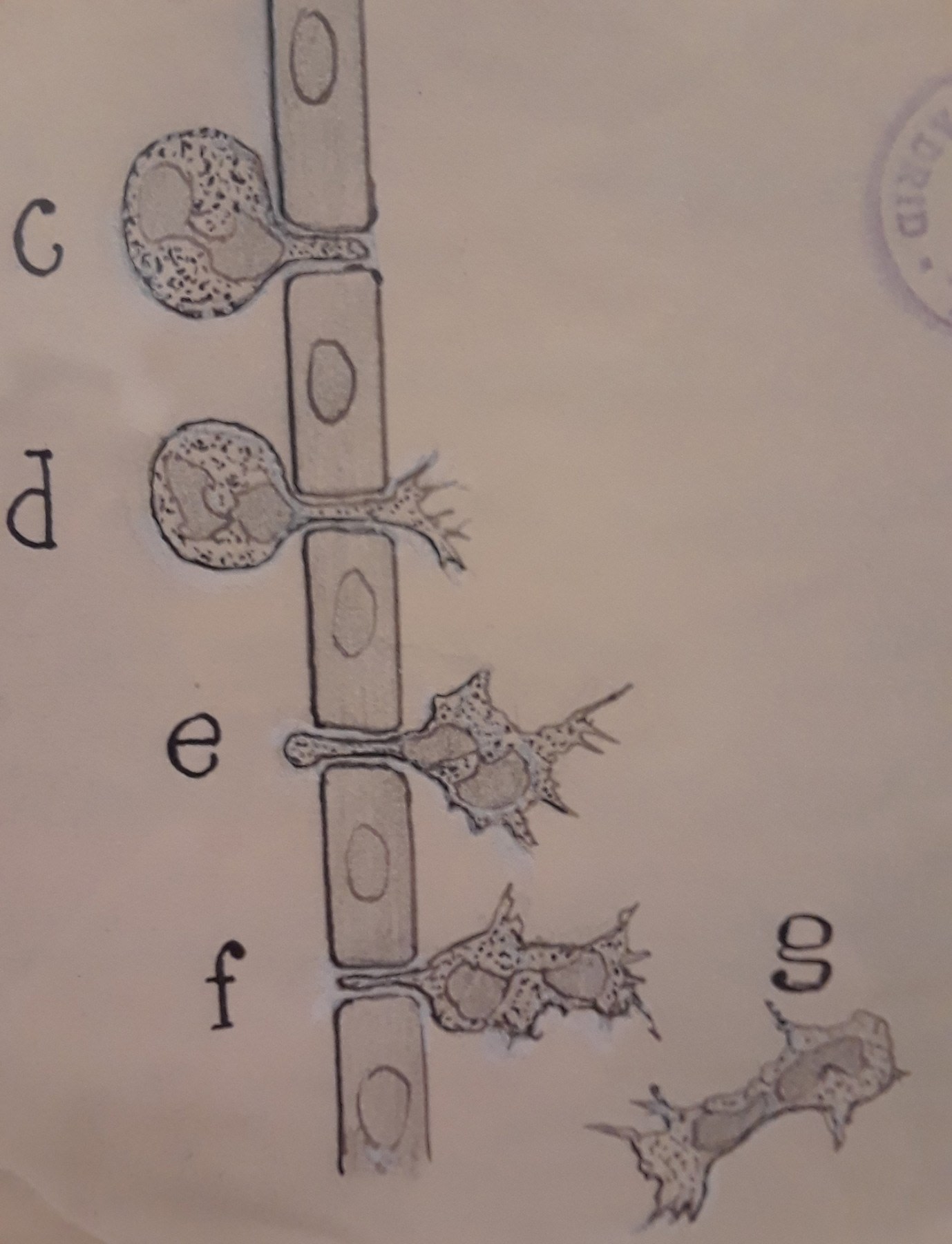
Figure 2 represents, in a sequential and didactic manner, the biological phenomenon known as leukocyte extravasation or diapedesis. This is a fundamental cellular process in the acute inflammatory response, by which leukocytes (white blood cells) migrate from the bloodstream, through the walls of the blood capillaries, into the surrounding tissues to combat pathogens or respond to injury. The sequence, marked with the letters c, d, e, f, and g, breaks down this complex event into comprehensible phases:
Figure 2: Extravasation of a Leukocyte
-
Phase (c): Shows a leukocyte, recognizable by its granular cytoplasm and amoeboid shape, in the margination and adhesion phase. The cell has moved toward the periphery of the blood flow and has adhered to the inner wall of the capillary, which is represented by the vertical structure of endothelial cells.
-
Phase (d): The leukocyte begins the transmigration phase. It extends a pseudopod (a cytoplasmic projection) that penetrates the junction between two adjacent endothelial cells, initiating its passage through the vascular barrier.
-
Phases (e) and (f): Illustrate the active passage of the leukocyte through the vessel wall. The cell demonstrates its remarkable plasticity and amoeboid motility, contracting and deforming to squeeze through the narrow intercellular space.
-
Phase (g): The leukocyte has completed extravasation. It is now completely outside the blood vessel, in the perivascular tissue, ready to migrate by chemotaxis toward the focus of inflammation.
1.3: A Visual Autopsy of Cellular Transmigration
A detailed analysis of his drawings on diapedesis reveals a sequential narrative that illustrates a dynamic process:
-
Initial Adhesion: A leukocyte of rounded and amoeboid morphology establishes initial contact with the inner wall of the blood vessel.
-
Probing and Polarization: The cell changes shape, extending a pseudopod toward the junction between two endothelial cells, demonstrating active and exploratory behavior.
-
Diapedesis - The Passage: In the critical stage, the cell contracts dramatically, squeezing its cytoplasm and nucleus through the narrow interendothelial space.
-
Completion and Migration: The greater part of the cell crosses the barrier, retracts its posterior end (uropod), and detaches completely from the vessel to migrate through the interstitial tissue, with a morphology fully adapted for amoeboid movement.
This act of editorial focus, isolating the key actors, is a hallmark of great scientific illustration. The drawing is not a literal copy but a conceptual model that communicates a mechanism.
1.4: The Molecular Choreography of Diapedesis
What Cajal observed morphologically is the final act of an exquisitely regulated molecular drama, known today as the leukocyte adhesion cascade.
-
Chemoattraction and Rolling: Cells from damaged tissue release cytokines (such as IL-1 and TNF-alpha) and chemokines, which activate endothelial cells. These express on their surface selectins, adhesion molecules that bind with low affinity to leukocytes, slowing them down and causing them to “roll” along the vessel wall.
-
Firm Adhesion: As they roll, leukocytes activate a family of receptors called integrins (such as LFA-1), which switch to a high-affinity conformation. These bind strongly to their ligands on the endothelium (such as ICAM-1), bringing the leukocyte to a complete halt.
-
Transmigration (Diapedesis): The physical passage through the barrier, predominantly between endothelial cells (paracellular route), is mediated by junctional molecules such as PECAM-1 and JAMs. This process is a cooperative dialogue, not a violent breach; endothelial cells actively form “transmigratory cups” to guide the leukocyte.
This first foray into cellular pathology was the crucible in which his scientific paradigm was forged. By observing the leukocyte as a discrete, mobile, and functional unit, Cajal was witnessing the quintessence of cellular individuality, the very same principle that, years later, would define his neuron doctrine.
Figure 3: The Immune Army on the Battlefield
Chapter 2: The Manual of Pathological Anatomy as a Hematological Codex
2.1: A Foundational and Evolving Work
In 1890, Cajal published the first edition of his Manual of General Pathological Anatomy, a work destined to become one of the most influential medical texts in the history of Spain. This manual, continuously updated over more than seven decades, trained entire generations of physicians.
2.2: Blood in Inflammation and General Pathology
Within the Manual, Cajal settled the debate on the origin of pus, writing that Cohnheim’s diapedesis “is today fully confirmed” (“esta hoy plenamente confirmada”). He conceptualized phagocytosis not as an act of war, but as a universal biological mechanism. In his own words, the phagocytic phenomenon:
… is more general than inflammation; it presents itself in the absorption of necrotic foci, in the atrophy and disappearance of embryonic organs (absorption of the tadpole’s tail, etc.), and represents the habitual means used by the organism to imprison carbon particles that penetrate the lung. (”… es mas general que la inflamacion; presentase en la absorcion de focos necroticos, en la atrofia y desaparicion de organos embrionarios (absorcion de la cola del renacuajo, etc.), y representa el medio habitual utilizado por el organismo para encarcelar las particulas de carbon que penetran en el pulmon.”)
2.3: The “Defensive Reaction” and the “Atavistic” Cell - Cajal as a Pioneer of Cancer Immunopathology
Perhaps the most prescient contribution of the Manual lies in the field of oncology. Cajal articulated a vision of the interaction between tumor and host that anticipated modern concepts by nearly a century. He described the cancer cell with astonishing lucidity:
The epithelioma cell is an anarchic cell, emancipated from the laws of the organism, which tends to live on its own account, like a parasite, and to reproduce not the organ to which it belonged, but an imperfect, atavistic cell type that recalls in a certain way the embryonic phases of the epithelium. (“La celula del epitelioma es una celula anarquica, emancipada de las leyes del organismo, que tiende a vivir por su cuenta, como un parasito, y a reproducir, no el organo a que pertenecio, sino un tipo celular imperfecto, atavico, que recuerda en cierto modo las fases embrionarias del epitelio.”)
In 1896, he proposed that the stroma surrounding the tumor was not mere scaffolding, but the stage of an active “defensive reaction” (“reaccion defensiva”) by the organism. He identified the combatants with precision: “lymphocytes, cells with chromophilic protoplasm, and Ehrlich’s corpuscles or Mastzellen” (“linfocitos, celulas con protoplasma cromofilo y corpusculos de Ehrlich o Mastzellen”). This was one of the earliest descriptions of an antitumor immune response. Going further, he intuited the possibility of immunotherapy, speculating on the need to find “substances capable of intensifying” (“sustancias capaces de exaltar la intensidad”) this defense.
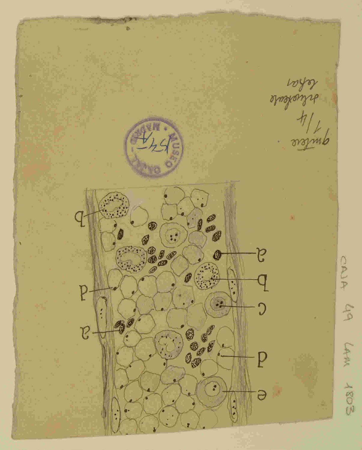
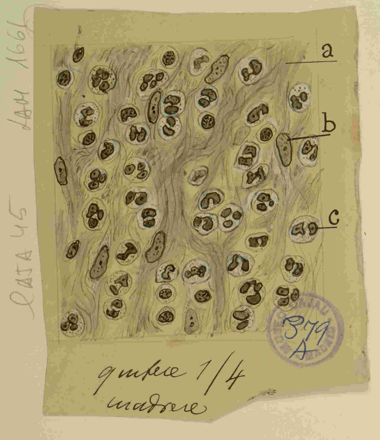
Figure 4: The Defensive Reaction
In the illustrations of a carcinoma, Cajal does not limit himself to drawing the tumor cells but represents the entire ecosystem: the reactive stroma, the blood vessels, and, crucially, the infiltration of immune cells. It is the direct visualization of his concept of “defensive reaction” and a portrait of the tumor microenvironment a century before its formal definition.
Chapter 3: Specific Investigations on Blood Corpuscles
3.1: The Enigma of the “Third Corpuscle”
At the end of the nineteenth century, the nature of platelets was the subject of an intense debate. While Georges Hayem considered them “hematoblasts” (precursors of red blood cells), Giulio Bizzozero correctly established their role in hemostasis.
3.2: “The Phagocytosis of Platelets” (1896)
In this context, Cajal published “The Phagocytosis of Platelets” (“La fagocitosis de las plaquetas”), in which he proposed that these cells possessed phagocytic capacity. This conclusion, now considered incorrect, reveals the force of his dominant paradigm: the search for cellular defense mechanisms. Having established the phagocytic role of leukocytes, it is logical that he would attempt to apply this model to other elements of the blood. This episode demonstrates both the strength of his systemic thinking and the limitations of any conceptual framework.
Chapter 4: An Essential Counterpoint - Cajal versus Paul Ehrlich, the Architect of Hematology
To understand Cajal’s place in hematology, it is indispensable to contrast him with his contemporary Paul Ehrlich (1854-1915), the founder of the discipline.
4.1: Paul Ehrlich - The Power of Chemistry
Ehrlich’s approach was revolutionary because it was grounded in chemistry. He developed techniques for staining blood smears with aniline dyes, which allowed him to observe that the different types of leukocytes possessed granules with different tinctorial affinities. Based on this “selective affinity,” he classified them into neutrophils, basophils, and eosinophils, a taxonomy that remains the cornerstone of the modern complete blood count.
4.2: Cajal - The Power of Morphology
Cajal’s paradigm was morphological. His principal tool was the Golgi silver impregnation method, which he perfected to reveal the complete three-dimensional structure of the cell and its connections. While Ehrlich sought to differentiate parts of the cell, Cajal sought to visualize the entire cell in its context.
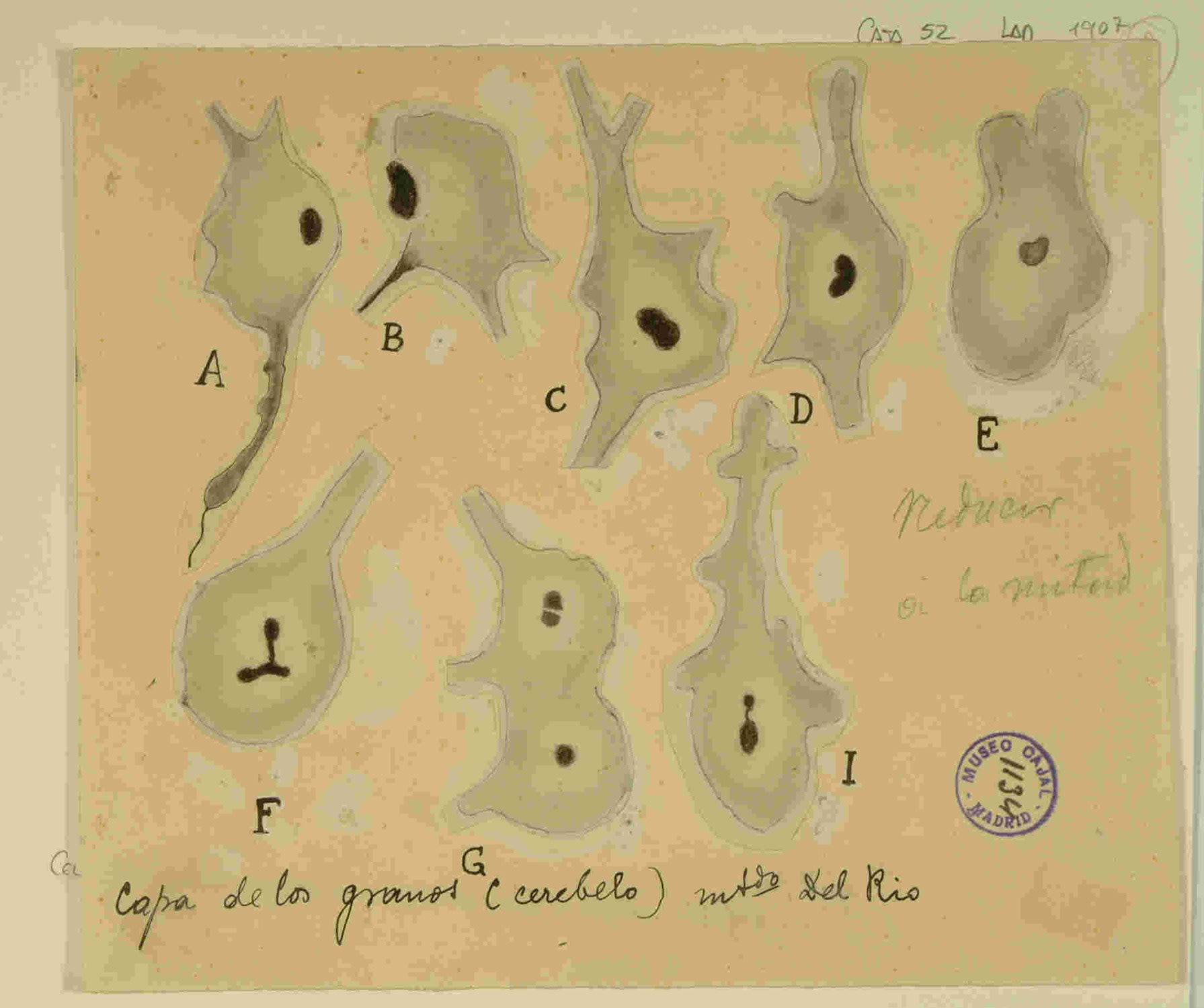
Figures 5 and 6: The Protagonists of Immunity
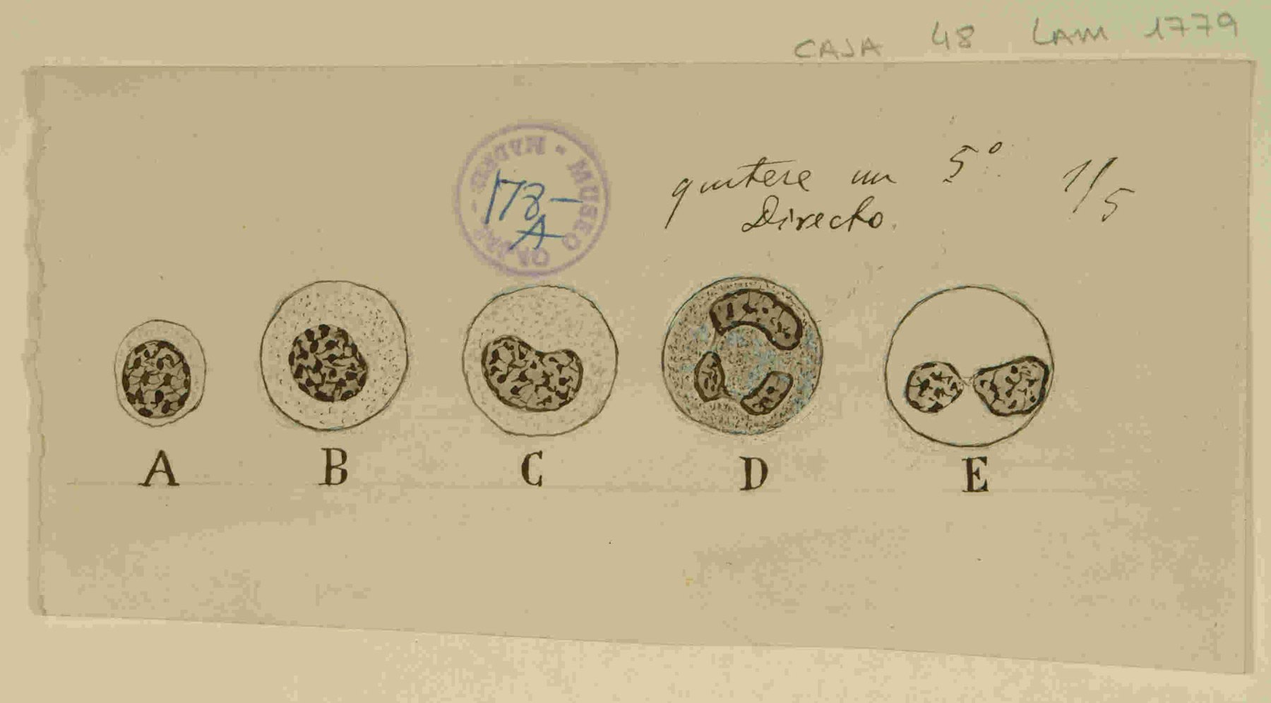
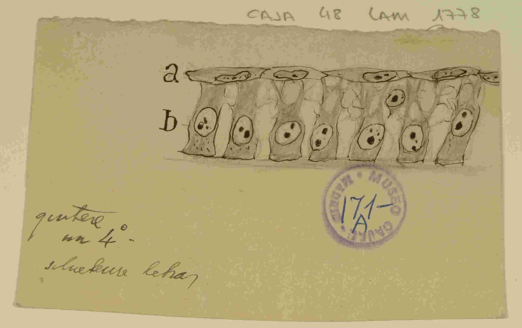
Table 1: Cajal vs. Ehrlich - Two Paradigms in the Study of the Blood Cell
| Characteristic | Santiago Ramon y Cajal / Paul Ehrlich / Main Field |
|---|---|
| Methodological Approach | Morphological-Structural (“See the complete form”)Chemical-Differential (“See the components”) |
| Key Techniques | Chromoargentic impregnation (Golgi method), Reduced silver nitrateTinctions with aniline derivatives, trichrome staining |
| Primary Object of Study | The cell in situ, within the tissueThe isolated cell (in blood smears) |
| Principal Contribution to Hematology | Description of cellular dynamics (diapedesis), characterization of the immune response in tissues (immunopathology), morphological studies of leukocytes and platelets.Classification of leukocytes (neutrophils, eosinophils, basophils), description of megaloblasts, foundation of hematology as a diagnostic discipline. |
| Legacy in the Field | Conceptual and functional: laid the foundations for immunopathology and the understanding of the cell as a dynamic agent.Foundational and taxonomic: created the tools and classification that define modern hematology. |
| Metaphor for His Contribution | The “Biographer” of the cell: narrated its behavior and its life within the tissueThe “Chemist” of the cell: revealed its internal composition and classified it |
| Export to Spreadsheets |
4.3: Cajal and Ehrlich - Modern Science Needs Both
Cajal and Ehrlich were not rivals, but the architects of two complementary scientific languages, both indispensable for describing the complex world of the blood. Ehrlich provided the “dictionary,” the precise taxonomy and classification of cell types that allowed hematology to become a rigorous clinical discipline. Cajal, for his part, wrote the “grammar,” the rules describing how those cells act, move, and interact within the organism. A dictionary without grammar is a mere list of terms; a grammar without a defined vocabulary cannot express complex ideas. Modern science needs both. This perspective resolves any debate about the primacy of one over the other and replaces it with a more sophisticated understanding of scientific progress, which advances through parallel methodological and conceptual revolutions.
Chapter 5: The Methodological Legacy: The Influence of the School of Cajal
5.1: Continuity through the Disciples
Cajal’s interest in pathology was perpetuated through the “School of Cajal.” Disciples such as Jorge Francisco Tello kept the legacy alive, updating his influential manuals.
5.2: Pio del Rio Hortega and Neuroimmunology
Pio del Rio Hortega (1882-1945) identified and characterized the “third element” of the nervous system, which he named microglia. He demonstrated that it was a cell of mesodermal origin, related to blood cells, and endowed with an extraordinary plasticity and phagocytic capacity. The same functional paradigm that Cajal had observed in leukocytes - motility, response to injury, and phagocytosis - was what Rio Hortega discovered in the brain’s resident macrophage, laying the foundations of neuroimmunology.
Figure 7: The Legacy of the School of Cajal
5.3: Pedro Ramon y Cajal - The Pioneer of Clinical Oncology
Santiago’s brother, Pedro (1854-1950), brought the rigor of the laboratory to clinical practice. He was a pioneer in the use of biopsy and radiotherapy in Spain, and one of the first to describe the phenomenon of radioresistance, anticipating key concepts such as clonal selection and acquired resistance.
Chapter 6: The Echo of Cajal in Twenty-First-Century Oncology
Cajal’s observations constitute the theoretical scaffolding upon which some of the most revolutionary advances in modern medicine have been built.
6.1: The “Germinal Corpuscles” and the Cancer Stem Cell (CSC) Theory
In 1896, Cajal proposed that tissues are composed of mature cells and “germinal corpuscles” (“corpusculos germinales”), described as “undifferentiated cells… that control tissue regeneration… and pathological processes.” He argued that tumors arose from these corpuscles. This concept is a direct precursor of the modern Cancer Stem Cell (CSC) theory, which postulates that tumors are maintained by a small subpopulation of cells with stem cell properties, responsible for tumor initiation, resistance, and relapse.
Figure 8: The Legacy of the School of Cajal
6.2: “Mitogenic Substances” and Anti-Angiogenic Therapy
In his Manual, Cajal described how a tumor cell “secretes mitogenic substances that act on the surrounding capillaries” (“segrega sustancias mitogenicas que actuan en los capilares… circundantes”). This is a perfect description of tumor angiogenesis, a conceptual precursor of the discovery of factors such as VEGF and of anti-angiogenic therapies that block this process. His genius lay in understanding angiogenesis not only as a pathological phenomenon but as a fundamental biological principle for tissue repair, as he illustrated in his works on the “restoration of circulation.”
6.3: The “Defensive Reaction” and the Era of Immunotherapy
Cajal’s idea of the stroma as a “defensive reaction” (“reaccion defensiva”) is the precursor of the concept of the Tumor Microenvironment (TME). His intuition about “intensifying” (“exaltar la intensidad”) this defense is the guiding principle of modern immunotherapy.
-
Immune Checkpoint Inhibitors: The immune system possesses “brakes” (such as PD-1 and CTLA-4). Many tumors exploit these mechanisms to “switch off” T lymphocytes. Checkpoint inhibitors are antibodies that block these interactions, releasing the brakes and allowing T lymphocytes to attack the tumor.
-
CAR-T Cell Therapy: This consists of genetically modifying a patient’s T lymphocytes so that they express a Chimeric Antigen Receptor (CAR) that recognizes a specific antigen on tumor cells, converting them into “guided missiles.”
6.4: Diapedesis as a Tragic Paradigm of Metastasis
The imitation of leukocyte extravasation by cancer cells is an evolutionary masterpiece of pathology. Cancer co-opts a host defense system for its own dissemination. Circulating Tumor Cells (CTCs) use similar mechanisms of adhesion (selectins, integrins) and chemotaxis to exit the vasculature. However, there is a critical divergence:
-
Leukocyte Diapedesis: A cooperative, temporary, and reversible process that maintains the integrity of the endothelium.
-
Tumor Extravasation: An often destructive and irreversible process that can permanently damage the vascular barrier, reflecting the invasive nature of cancer.
Cajal’s drawing of the “loyal soldier” (the leukocyte) thus becomes a tragic template for the “traitor” (the cancer cell).
Chapter 7: The Drama of Circulation: Thrombi, Emboli, and Repair
Cajal’s genius as a pathologist was not limited to inflammation or cancer; it encompassed an integral vision of vascular dynamics. His drawings from the Cajal Legacy reveal a profound understanding of the processes that govern both disease and the repair of blood vessels. In works such as “Mechanism of the Hemorrhagic Infarction” (“Mecanismo del infarto hemorragico”) and “Course of the Embolus” (“Marcha del embolo”), he unraveled with his ink scalpel the mechanisms of thrombosis and embolism, illustrating the destructive consequences of vascular obstruction.
However, his vision did not stop at pathology. As a counterpart, he studied and drew the “Mechanism for the Restoration of Circulation in Organic Territories” (“Mecanismo del restablecimiento de la circulacion en los territorios organicos”). This work is definitive proof that he understood angiogenesis not only as a pathological process but as a fundamental biological principle for repair and life. This duality in his thinking is extraordinary: he was able to see the same process - the formation of new vessels - as a healing mechanism in damaged tissues and as the engine of growth in tumors, anticipating the modern idea that cancer hijacks and perverts the normal functions of the organism for its own benefit.
Conclusion: Redefining the Hematological Legacy of Santiago Ramon y Cajal
The vast shadow cast by the neuroscientific legacy of Santiago Ramon y Cajal has kept in the penumbra a set of contributions that, analyzed in their historical and scientific context, prove to be fundamental and extraordinarily prescient. An exhaustive analysis of his work reveals that, far from being a field unrelated to his interests, blood pathology and the immune response were central themes in the development of his biological thinking.
The synthesis of his contributions paints the profile of a scientist of astonishing coherence. In the field of inflammation, his verification of diapedesis established his paradigm of the cell as an individual agent. In vascular pathology, he unraveled the mechanisms of thrombosis, embolism, and reparative angiogenesis. But it is in oncology where his “other doctrine” reveals his most visionary genius. His concepts of the “atavistic cell” (“celula atavica”), the “germinal corpuscles” (“corpusculos germinales”), and “mitogenic substances” (“sustancias mitogenicas”) anticipated, respectively, tumor de-differentiation, the cancer stem cell theory, and angiogenesis. His description of the stroma as a “defensive reaction” (“reaccion defensiva”) and his intuition about how to “intensify” this response laid the conceptual foundations for immuno-oncology and modern immunotherapy.
Figure 9: The Scientist and His Tool
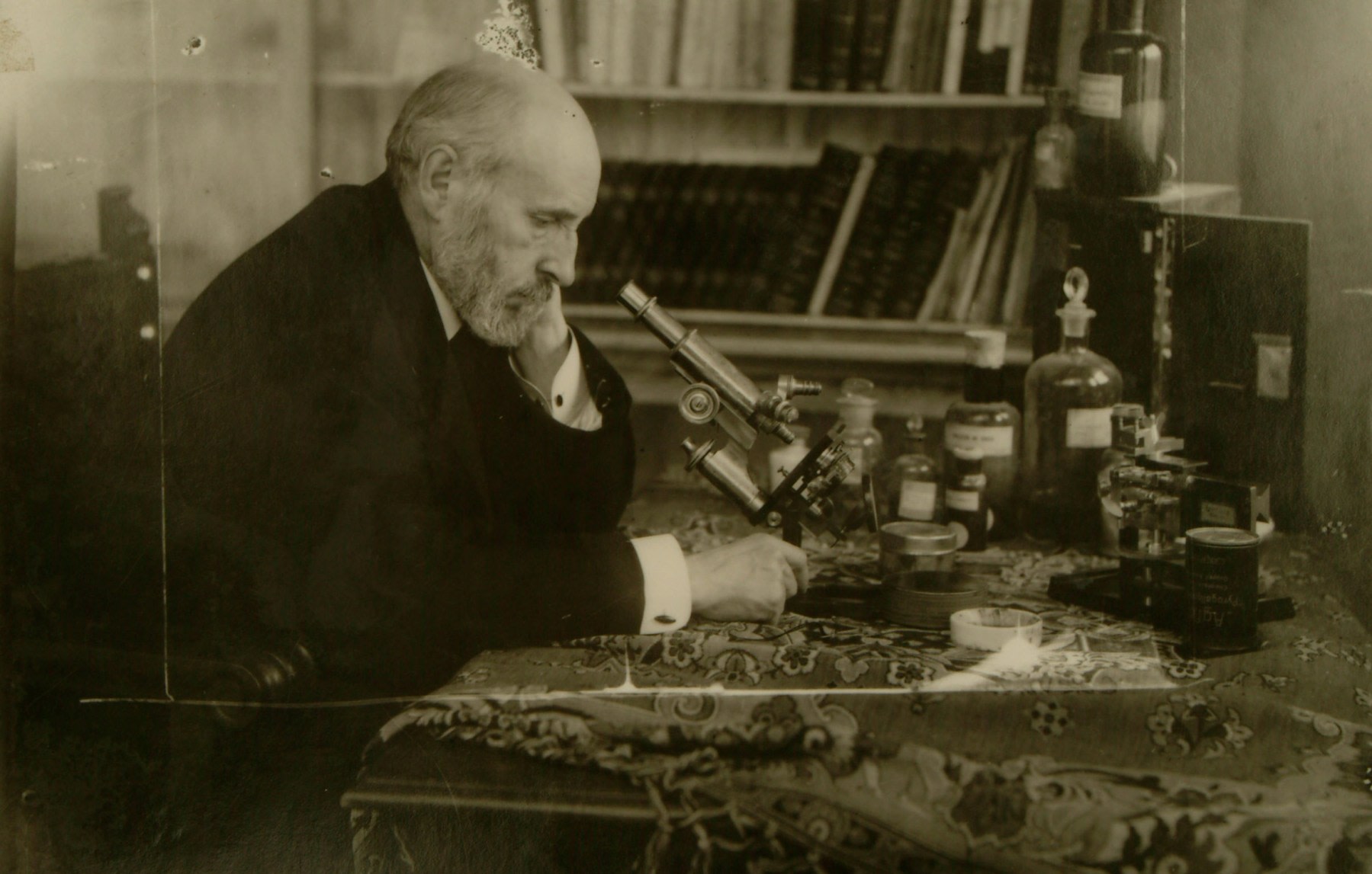
In the final evaluation of his legacy, it is evident that the title of “father of hematology” belongs to Paul Ehrlich. Cajal, on the other hand, was not a classifier but a cell biologist who deciphered the function of cells in the drama of disease. For this reason, he must be recognized as a figure of the first order in the history of cellular pathology and an indisputable pioneer of modern oncology. Cajal always placed the cell as the protagonist of life, whether in the intricate networks of the brain or in the inflammatory battle of a blood vessel.
His work, in its entirety, compels us to broaden our perspective and to recognize him not only as the cartographer of the brain, but as a universal explorer of the “enchantment of the infinitely small” (“hechizo de lo infinitamente pequeno”), whose forays into the bloodstream and tumor tissues revealed truths as profound and enduring as those he found among the “mysterious butterflies of the soul” (“misteriosas mariposas del alma”).
Bibliography
-
Ramon y Cajal, S. (1880). Investigaciones experimentales sobre la genesis inflamatoria y en especial sobre la emigracion leucocitaria. Zaragoza: Imprenta El Diario Catolico.
-
Ramon y Cajal, S. (1896). Manual de Anatomia Patologica General (2nd ed.). Madrid: Imprenta y Libreria de Nicolas Moya.
-
Ramon y Cajal, S. (1896). La fagocitosis de las plaquetas. Revista Trimestral Micrografica, 1(4).
-
Garcia-Lopez, P., Garcia-Marin, V., & Freire, M. (2005). The contributions of Santiago Ramon y Cajal to cancer research - 100 years on. Nature Reviews Cancer, 5(11), 904-909.
-
Rio-Hortega, P. del. (1919). El ‘tercer elemento’ de los centros nerviosos. Boletin de la Sociedad Espanola de Biologia, 9, 68-120.
-
Sanchez-Garcia, I., & Cobaleda, C. (2024). Childhood leukemia prevention within reach. Blood, 144(8), 799-800.
-
Waldman, A. D., Fritz, J. M., & Lenardo, M. J. (2020). A guide to cancer immunotherapy: from T cell basic science to clinical practice. Nature Reviews Immunology, 20(11), 651-668.