In the pages of his monumental work, Santiago Ramón y Cajal described the brain as an “impenetrable forest” where the scientific spirit ran the risk of losing itself amid the tangle of silver threads. Today, more than a century after his ink drawings defined the “Neuron Doctrine,” neuroscience has reached a milestone that resonates as a fulfilled prophecy: the cartography of 1 cubic millimetre of human temporal cortex at nanometric resolution. What to the naked eye appears as a fragment of tissue smaller than a grain of rice conceals within it a galaxy.
This project, called H01, is not merely a triumph of data engineering; it is the direct heir to Cajal’s gaze, now empowered by scanning electron microscopy and Artificial Intelligence algorithms that have enabled the “stitching” of biological reality into a coherent volume. The scale is overwhelming: a similar reconstruction for an entire human brain would require 1.6 zettabytes of storage, a capacity that today would demand the planet’s largest data centre.
The journey to the invisible: 326 days of capture
The acquisition of this petavoxel atlas began with a sample from the middle temporal gyrus, extracted during a neurosurgical intervention to treat epilepsy in a 45-year-old woman. To preserve the “architecture of thought” before post-mortem degradation erased it, the tissue was subjected to immediate fixation, achieving an ultrastructural fidelity that Cajal could only have intuited in his preparations.
The imaging process was a technical odyssey of 326 uninterrupted days of operation. The sample was sliced into 5,000 sections, each with a thickness of barely 30 nanometres—orders of magnitude thinner than a human hair. The result is a three-dimensional network in which 57,000 cells, 230 millimetres of blood vessels, and an overwhelming census of 150 million synapses have been identified.
| Attribute of the micro-universe | Quantitative value |
|---|---|
| Fragment volume | 1.05 mm³ |
| Image acquisition time | 326 days |
| Data generated | 1.4 Petabytes (1,400 Terabytes) |
| Total cells (neurons and glia) | 57,180 |
| Blood vessel length | 230 mm |
| Automatically annotated synapses | ~150 million |
AI: the new microscope of the Spanish School
The reconstruction of each neuron and each cellular process within this ocean of data was made possible through the collaboration with Google Research and the use of Flood-Filling Networks (FFN). This use of Artificial Intelligence closes a fascinating historical circle: the architecture of modern artificial neural networks is inspired by the principles of distributed processing that Cajal began to delineate.
Furthermore, the legacy of Rafael Lorente de Nó, Cajal’s most beloved disciple, resonates in every algorithm. Lorente de Nó proposed “reverberating circuits,” feedback loops that freed the brain from the “tyranny of the immediate stimulus.” This concept is the conceptual basis of the recurrent neural networks that today enable machines to process language and, in this study, to reconstruct the morphology of neurons with an unprecedented precision.
Cellular geography: dismantling the myth of glia
One of the most revealing results of the H01 study is the confirmation that, in the human cerebral cortex, glia is the silent majority, outnumbering neurons in a ratio of 2:1. This finding puts an end to decades of debate about the glia-neuron ratio, which oscillated between mythical estimates of 10:1 and more recent counts of 1:1.
Within this ecosystem, oligodendrocytes are the most abundant cells, distributed in a gradient that increases toward the white matter. Astrocytes, for their part, are organised in a mosaic of territorial domains, a configuration that guarantees the metabolic support necessary for synaptic activity.
The mirror symmetry of layer 6: compass neurons
In the depths of layer 6, researchers discovered a class of cells called “triangular” or “compass” neurons that possess an unexpected geometric order. Unlike conventional pyramidal neurons, these possess a massive basal dendrite that is not randomly oriented.
Analysis revealed that these neurons organise themselves in pairs with a “mirror symmetry”: while some direct their basal dendrites in one direction, their neighbours tend to do so in the opposite direction with a statistically significant probability of P = 0.005. This structural sophistication suggests the existence of specialised circuits for large-scale signal integration, a dimension that adds depth to the classical cytoarchitecture described by the sage.
Defying chance: connections of extreme power
The most disruptive discovery of H01 is the existence of multi-synaptic connections of unusual strength. The traditional view, based on “Peters’ Rule,” suggests that neuronal connectivity is largely stochastic.
However, petavoxel data show that the human brain is not governed solely by chance. While the vast majority of connections are weak, exceptional cases were found where a single axon establishes up to 50 synapses with a single postsynaptic neuron. These axons appear to actively “seek” their partners, wrapping around the target dendrite to ensure maximum fidelity signal transmission. These “high-speed bridges” could be the physical foundations of our most stable memories.
| Type of synaptic link | Frequency | Probable nature |
|---|---|---|
| Single synapse | 96.49% | Incidental or plastic connections |
| 3-synapse connections | 0.35% | Uncommon specialised circuits |
| Multi-synapse (up to 50) | Exceptional | High-fidelity functional “anchors” |
The enigmas of the tissue: whorls and anomalies
Nanometric resolution has also revealed “uncomfortable secrets” in the tissue. So-called “axonal whorls” have been documented—knots where axons wind extensively upon themselves forming spirals of membranous material. Since the sample comes from a patient with refractory epilepsy, the mystery remains: are these knots scars of the disease, effects of pharmacotherapy, or normal but extremely rare features of human nature? This uncertainty underscores the need to continue this atlas across diverse samples to define, with the precision that the sage demanded, the biological norm.
Epilogue: the future of engramics
The H01 fragment is the most detailed map ever created of human structure, a “Genome of Connectivity” that opens the door to the era of “engramics”: the study of how physical experience is translated into neuronal wiring. By sharing this resource openly, the researchers follow Cajal’s intellectual generosity, allowing the global scientific community to scrutinise each petavoxel in search of the spark of consciousness.
From the grain of sand to the zettabyte horizon, the indomitable spirit of Santiago Ramón y Cajal continues to guide our hand. As he himself warned: “As long as our brain remains a mystery, the universe, a reflection of the brain’s structure, will also be a mystery” (“Mientras nuestro cerebro sea un misterio, el universo, reflejo de la estructura del cerebro, también será un misterio”). With this atlas, the mystery has at last begun to yield to the light of reason.
Image credits
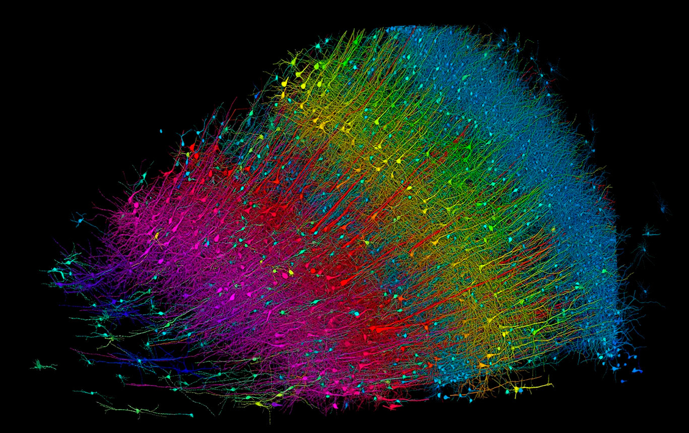 Three-dimensional reconstruction of the six cortical layers of the human temporal cortex, with excitatory neurons coloured by depth.
Credits: Google Research and Lichtman Lab (Harvard University).
Rendering: D. Berger (Harvard University).
Three-dimensional reconstruction of the six cortical layers of the human temporal cortex, with excitatory neurons coloured by depth.
Credits: Google Research and Lichtman Lab (Harvard University).
Rendering: D. Berger (Harvard University).
Recommended bibliography
-
Shapson-Coe, A., Januszewski, M., Berger, D. R., et al. (2024). A petavoxel fragment of human cerebral cortex reconstructed at nanoscale resolution. Science, 384(6696), eadk4858. DOI: 10.1126/science.eadk4858
-
Januszewski, M., Kornfeld, J., Li, P. H., & Jain, V. (2018). High-precision automated reconstruction of neurons with flood-filling networks. Nature Methods, 15(8), 605-610. DOI: 10.1038/s41592-018-0049-4
-
Loomba, S., Straehle, J., Gangadharan, V., et al. (2022). Connectomic comparison of mouse and human cortex. Science, 377(6601), eabo0924. DOI: 10.1126/science.abo0924
-
Lorente de Nó, R. (1938). Analysis of the activity of the chains of internuncial neurons. Journal of Neurophysiology, 1(3), 207-244. DOI: 10.1152/jn.1938.1.3.207
-
Ramón y Cajal, S. (1904). Textura del sistema nervioso del hombre y de los vertebrados. Imprenta de Nicolás Moya, Madrid.